

GCSE
Biology
-
Introduction to GCSE Biology (AQA) Coming soon
-
1.1 Cell Structure
-
1.2 Cell Division Coming soon
-
1.3 Transport in Cells Coming soon
-
2.1 Principles of Organisation Coming soon
-
2.2 Animal Tissues, Organs and Organ Systems Coming soon
-
2.3 Plant Tissues, Organs and Systems Coming soon
-
3.1 Communicable Diseases Coming soon
-
3.2 Monoclonal Antibodies [HT] Coming soon
-
3.3 Plant Disease Coming soon
-
4.1 Photosynthesis Coming soon
-
4.2 Respiration Coming soon
-
5.1 Homeostasis Coming soon
-
5.2 The Human Nervous System Coming soon
-
5.3 Hormonal Coordination in Humans Coming soon
-
5.4 Plant Hormones Coming soon
-
6.1 Reproduction Coming soon
-
6.2 Variation and Evolution Coming soon
-
6.3 The Development of Understanding of Genetics and Evolution Coming soon
-
6.4 Classification of Living Organisms Coming soon
-
7.1 Adaptations, Interdependence and Competition Coming soon
-
7.2 Organisation of an Ecosystem Coming soon
-
7.3 Biodiversity and the Effect of Human Interaction on Ecosystems Coming soon
-
7.4 Trophic Levels in an Ecosystem Coming soon
-
7.5 Food Production Coming soon
1. Cell Biology
In this lesson, we will explore how microscopyThe use of microscopes to observe small structures. has developed over time and how improvements in technology have allowed scientists to study cells in increasing detail. We will compare light and electron microscopes in terms of magnificationThe number of times larger an image appears compared to the real object. and resolutionThe ability to distinguish two separate points as distinct., and practise calculations involving image sizeThe measured size of an object as seen under a microscope., real size and magnification, including expressing answers in standard formA way of writing very large or very small numbers as a number between 1 and 10 multiplied by a power of ten. where required.
Development of Microscopy Techniques
Microscopy has played a vital role in the development of biology. Cells are too small to be seen with the naked eye, so microscopes allow scientists to observe their structure and organisation. As microscopes have improved, our understanding of cells and sub-cellular structures has become much more detailed.
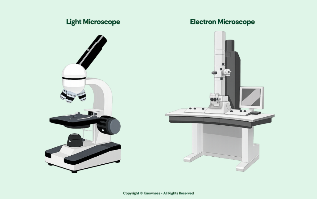
The first widely used microscopes were light microscopes. These use visible light and glass lenses to magnify specimens. For many years, light microscopes were the main tool for studying cells and tissues, and they are still widely used in schools and laboratories today.
In the 20th century, electron microscopes were developed. Instead of using light, they use a beam of electrons. Because electrons have a much shorter wavelength than visible light, electron microscopes can achieve much higher magnification and resolution. This has allowed scientists to observe much smaller structures inside cells.
Magnification and Resolution
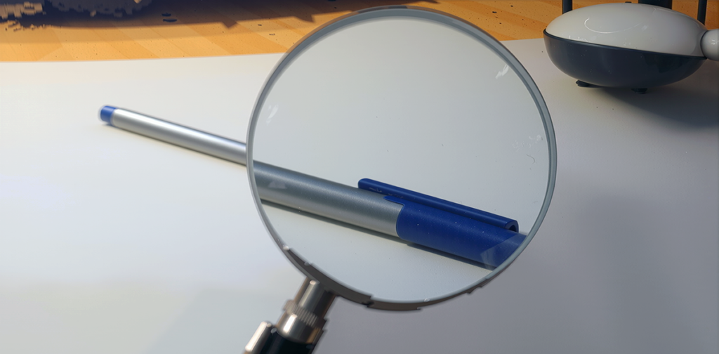
Magnification and resolution are two different but related concepts. Magnification describes how many times larger an image appears compared to the real object (i.e. the ratio of an object’s image size to its real size).
Resolution, on the other hand, is the ability to distinguish two separate points as distinct from each other (i.e. a measure of the clarity of the image).
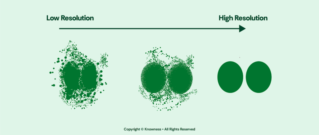
Higher magnification does not always mean clearer detail. Magnification makes an image larger, but resolution determines how clear it is. Increasing magnification without improving resolution only makes a blurry image bigger. Note
Light microscopes typically magnify up to about ×1000. Their maximum resolution is around 200 nanometres (nm). This means that if two structures are closer together than 200 nm, they will appear as one blurred structure. Very small structures such as ribosomes cannot be seen clearly using a light microscopeA microscope that uses visible light and glass lenses to magnify specimens..
A light microscope is like using binoculars. You can see large features clearly, but very fine details remain blurred because visible light has limits.Analogy
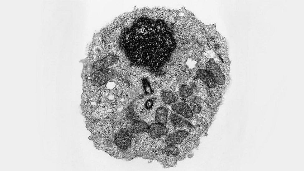
In comparison, electron microscopes can magnify images hundreds of thousands to over a million times. More importantly, they have a much higher resolution, down to a few nanometres. This allows scientists to observe very small sub-cellular structures, including ribosomes and internal membrane details within mitochondriaAn organelle where aerobic respiration occurs and energy is released. and chloroplasts. Because of this increased resolution, electron microscopy has greatly improved our understanding of cell structure and function.
An electron microscopeA microscope that uses a beam of electrons to produce highly magnified, high-resolution images. is like high-resolution satellite imaging. It reveals extremely fine detail that ordinary viewing methods cannot detect.Analogy
Magnification Calculations
When using microscopes, it is important to calculate magnification correctly so that the real size of structures can be determined. The formula for magnification is:
\(\text{Magnification} = \frac{\text{Size of Image}}{\text{Size of Real Object}}\)
To calculate magnification, you divide the image size by the real size of the object. The image size is how large the specimen appears under the microscope, while the real size is its actual dimension, usually much, much smaller.
Let’s say we have observed an image under the microscope, and the size of the image is 3 mm. The actual size of the object being viewed is 0.1 mm. \(\text{Magnification} = \frac{\text{Size of Image}}{\text{Size of Real Object}}\) \(\text{Magnification} = \frac{3\text{\, mm}}{0.1\text{\, mm}} = 30\) The units cancel, leaving magnification as a number without units. Since 30 is not very large or very small, it does not need to be written in standard form.Example
When performing microscopy calculations, if the result is very small or very large then standard form can be used. Some higher mark questions will ask for the answer in standard form, so double check the instructions.Tip
Magnification does not improve resolution. Electron microscopes provide greater detail mainly because of their higher resolution, not just because they magnify more.Note
Development of Microscopy Techniques
- Microscopy allows scientists to observe cells and sub-cellular structures that cannot be seen with the naked eye.
- Improvements in microscopes have increased our understanding of cell structure and organisation.
- Light microscopes use visible light and glass lenses to magnify specimens.
- Electron microscopes use a beam of electrons instead of light.
- Because electrons have a shorter wavelength than visible light, electron microscopes achieve much higher magnification and resolution.
- Electron microscopy allows scientists to observe much smaller structures inside cells.
Magnification and Resolution
- Magnification describes how many times larger an image appears compared to the real object.
- Resolution is the ability to distinguish two separate points as distinct from each other.
- Increasing magnification without improving resolution only makes a blurry image larger.
- Light microscopes typically magnify up to about ×1000.
- The maximum resolution of a light microscope is around 200 nanometres (nm).
- Structures closer together than 200 nm appear as one blurred structure under a light microscope.
- Electron microscopes can magnify images hundreds of thousands to over a million times.
- Electron microscopes have a much higher resolution than light microscopes, down to a few nanometres.
- Electron microscopes can reveal very small sub-cellular structures such as ribosomes and internal membrane details.
- The improved resolution of electron microscopes has greatly advanced understanding of cell structure and function.
Magnification Calculations
- Magnification is calculated using the formula: \(\text{Magnification} = \frac{\text{Size of Image}}{\text{Size of Real Object}}\).
- Magnification has no units because the units cancel during calculation.
- The image size is how large the specimen appears under the microscope.
- The real size is the actual size of the object.